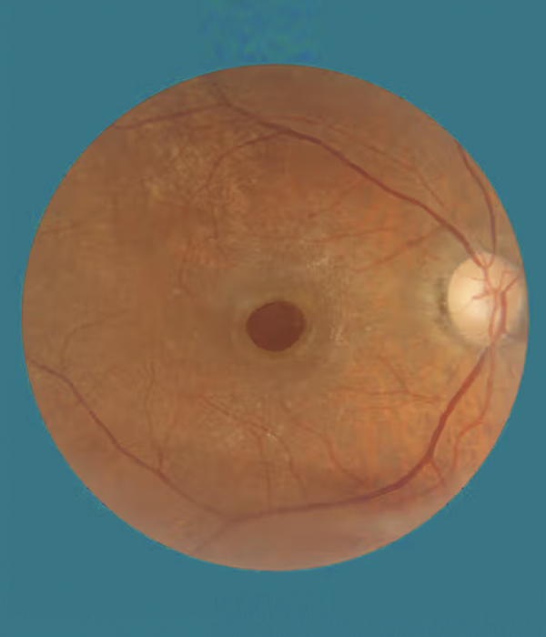
Epi retinal membrane (ERM)
Home / Retina / Epi retinal membrane
ERMs
ERMs can be diagnosed during a routine vision exam. In many cases, vision is not affected. Most types of ERM do not change and cause vision symptoms.
Some ERM do get worse, however, and will cause blurring and vision disturbances. The only time a doctor will suggest treatment is when there are vision symptoms.
A diagnostic test called optical coherence tomography (OCT) which uses light waves to scan and view the layers of the retina, can help with the diagnosis of ERMs.
We may also use another test called fluorescein angiography. This test involves the use of dye to light up areas of the retina.
About 15 percent of ERMs require surgery, this according to one report from the Indian Journal of Ophthalmology. Further, surgical intervention is successful in most cases, even though vision improvement for 25 to 50 percentb is at about 20/40.
The 20/40 measurement is used to define visual accuracy, clarity, and sharpness. A 20/40-vision measurement means someone sees at 20 feet (ft) what a person with normal vision would see at 40 ft.


Vitrectomy surgery procedure
The surgery for ERMs is called a vitrectomy. During a vitrectomy, the surgeon will make tiny cuts in the affected eye and remove the fluid from inside the eye.
The surgeon will then hold and gently peel the epiretinal membrane from the retina and replace the fluid in the eye.
Finally, the doctor places a pad and shield on the eye to protect it from infection or injury.

